Memorial Sloan Kettering Cancer Center

Deasy Laboratory for Computational
Medical Physics and
Mathematical Oncology

Physics and information based modeling, outcome prediction, and optimization of cancer treatment. Integrating dose, imaging, and molecular data to understand cancer and optimize treatments.

0Peer-reviewed papers
0Trainees mentored
0Patients treated (ECHO)
0Major NIH awards led
Single-cell p53–p21 dynamics after radiation Cell-to-cell variability in the p53 response over time — the kind of signal our models are built to read. Adapted from Tran et al., Cell Death & Differentiation (2023).

Why our work matters

Turning cancer's variability into better treatment

Cancer varies — from patient to patient, tumor to tumor, and cell to cell. The Deasy Lab treats that variability as information: we build models that read tumor biology from routinely acquired images, genomes, and clinical data, and turn it into decisions that make treatment more precise and less toxic. The aim is not modeling for its own sake, but tools that reach the clinic and change how patients are cared for.

Lab News — updated weekly Every paper, preprint, and news story from the last two years involving the PI or a current lab trainee.
Open the Lab News feed →
Mentee News — updated weekly High-profile papers, media, and awards from the lab's current and former trainees, with where each of them is now.
Open the Mentee News feed →

Research programs

Four connected lines of work

The through-line across all of them: innovative mathematics used to understand patient and disease variability — and to turn it into a leverage point for cancer care.

Mathematical Oncology Initiative

Geometry & optimal transport for the structure of cancer

Breast Cancer Research Foundation · Simons Foundation

Geometry- and topology-based methods to subtype patients, integrate multi-omic networks, and surface therapeutic targets — and to train the next generation who build them.

optimal transportnetwork curvatureNetFlow
ECHO

Automated, clinically optimal treatment planning

2021 INFORMS Franz Edelman Laureate

A prioritized optimization that turns the hand-tuned trade-off between tumor coverage and organ sparing into a reproducible, mathematically optimal plan — now used to help plan roughly 23% of patients treated at MSK.

Proton & particle radiobiology

Modeling response across radiation modalities

A thread since the lab's first NIH award

Mechanistic models of how dose, timing, and tumor biology combine to determine response and toxicity when the physics differs from conventional photons — reproducing clinical observations across photon and carbon-ion therapy.

Digital twins

A simulatable model of each patient's tumor

Multimodality imaging + tumor biology

Combining FDG-PET and MRI with a mechanistic tumor-control model to forecast response and flag likely non-responders early — while treatment can still be adapted.

Tools for cancer research

Software we build, use, and share

Open platforms and methods from the lab — for images and dose, for the geometry of omics data, for AI segmentation in the clinic, and for keeping a research program organized.

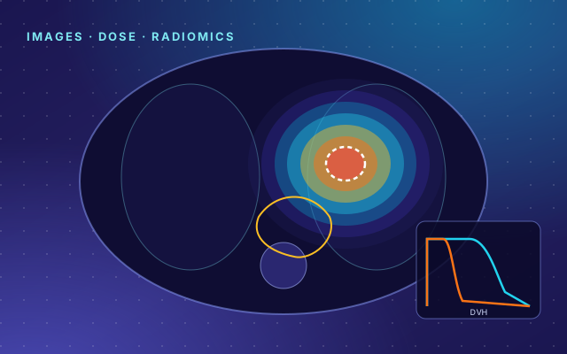
CERR / pyCERR: a CT slice with dose colorwash, contours and a dose-volume histogram

CERR / pyCERR

The lab's open-source platform for relating medical images to radiotherapy dose and outcomes — registration, dose calculation, contouring, and quantitative ("radiomic") image analysis. CERR has underpinned 70+ of the lab's studies and been cited over 800 times; pyCERR is its native Python successor, with a graphical viewer and AI-pipeline utilities. Development is led by Aditya Apte, PhD.

PythonMATLABOpen source

Apte et al., Medical Physics (2018), PMC6597320.

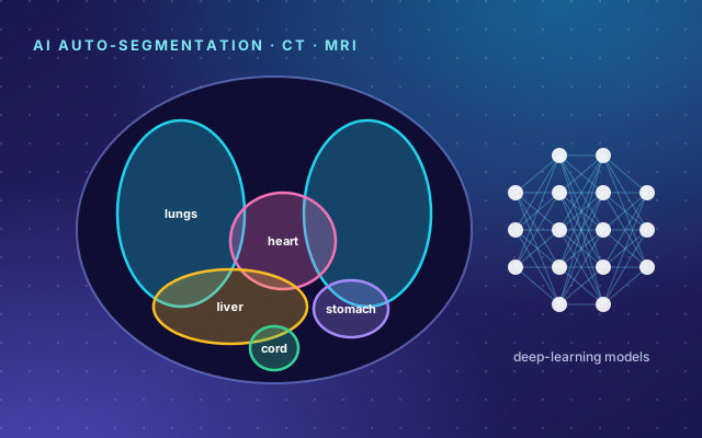
AI auto-segmentation: organs outlined in color on a torso cross-section beside a neural-network motif

AI auto-segmentation models

Deep-learning models for segmenting tumors and organs at risk on CT and MRI — heart and cardiac substructures, lung, head-and-neck, prostate, rectum, pancreas, and whole-body models — published with pyCERR and installable with one command via the CERR model installer. Models of this kind are in routine clinical use for radiotherapy planning at MSK.

CT & MRI12 published modelspyCERR
ORCO: a gene network whose edges are colored by Ollivier-Ricci curvature

ORCO

Ollivier-Ricci Curvature-Omics: an open-source Python package that lays omics data over gene- or protein-interaction networks and measures the curvature of each interaction — a geometric readout of how robust or fragile a biological system is, which can surface hidden therapeutic targets and regulatory modules. Install with pip install orcomics.

PythonNetwork geometryBioinformatics 2025
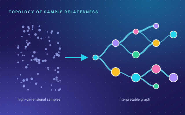
NetFlow: high-dimensional samples flowing into an interpretable branching graph

NetFlow

A framework for exploring topological representations of high-dimensional biomedical data. NetFlow turns a cohort of samples into an interpretable graph of sample relatedness — built on optimal transport, diffusion, and topological data analysis — to support precision oncology and hypothesis generation, with Jupyter tutorials that reproduce the multiple-myeloma analysis from the paper.

PythonTopology · OMTPreprint 2025
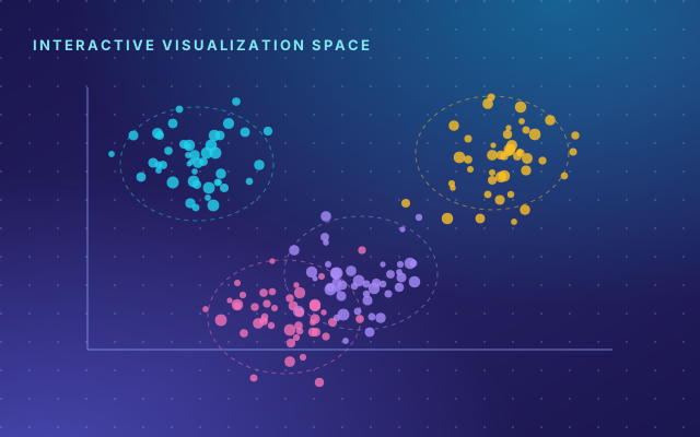
ViSPACE: clusters of tissue regions mapped across a whole-slide image

ViSPACE

Virchow2-powered Spatial Characterization and feature Extraction. An end-to-end computational pathology pipeline for whole-slide images of lung and breast cancer: it pairs the Virchow2 pathology foundation model with a pixel-wise decoder to segment tissue into tumor, stroma, inflammatory cells (TILs), necrosis and other, then extracts quantitative spatial biomarkers of the tumor microenvironment — tumor–stroma ratio, stromal TIL scores, immune and necrosis proximity, and tumor morphology. Developed by Himangi Srivastava; open source (Apache 2.0), paper under submission.

Computational pathologyPython

Recent work

Selected recent publications

A few highlights — the full, searchable library has every paper with a plain-language summary.

Imaging AI

Tumor hypoxia from a routine PET scan

An AI model estimates a head-and-neck tumor's oxygen-starved volume directly from routine FDG-PET — no specialized hypoxia scan required.

Physics and Imaging in Radiation Oncology, 2025
Language models

Predicting progression from clinical text

A large language model trained on oncology records extracts signals of cancer progression from free-text reports at scale.

npj Digital Medicine, 2025
Spatial modeling

A multi-institutional brain-metastasis atlas

Pooled imaging maps where brain metastases arise, informing precise detection and function-sparing radiotherapy.

Nature Communications, 2025

The lab

A team at the interface of mathematics and medicine

Led by Joseph O. Deasy, PhD, Chair of Medical Physics at MSK, the lab brings together mathematicians, physicists, and computational scientists.

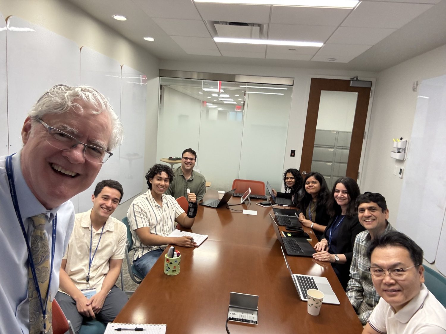
The Deasy Laboratory for Computational Medical Physics and Mathematical Oncology meeting around a conference table. Left to right: Joseph Deasy, PhD (PI); Daniel Grange, PhD (postdoc); Francisco Calderon (undergraduate); Anish Simhal, PhD (postdoc); Himangi Srivastava, PhD (postdoc); Lopamudra Nayak, PhD (postdoc); Leyla Ebrahimpour, PhD (postdoc); Aditya Apte, PhD (faculty); Jung Hun Oh, PhD (faculty).
Left to right: Joseph Deasy, PhD (PI); Daniel Grange, PhD (postdoc); Francisco Calderon (undergraduate); Anish Simhal, PhD (postdoc); Himangi Srivastava, PhD (postdoc); Lopamudra Nayak, PhD (postdoc); Leyla Ebrahimpour, PhD (postdoc); Aditya Apte, PhD (faculty); Jung Hun Oh, PhD (faculty).

Join us · Collaborate

We're recruiting — and we welcome collaborations

We welcome postdoctoral fellows, graduate students, and clinical fellows from mathematics, physics, computer science, engineering, statistics, and medicine — and research collaborations across imaging, genomics, and radiotherapy.